
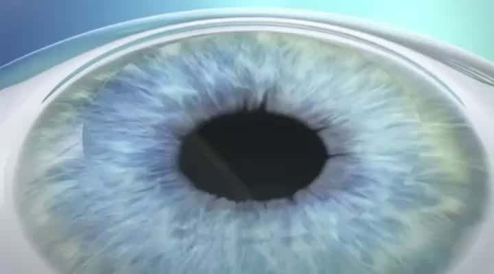
Glaucoma
Experience Clearer vision with advanced, personalized laser eye surgery tailored to your unique needs.
Glaucoma Brochures:
Download our glaucoma brochure to discover how the condition develops, which proven treatment options are available, and why thousands have trusted Re:Vision's specialists to protect their vision and reduce their reliance on daily eyedrops.


Why choose PRK ?
What is LASIK ?
Why choose ICL ?
- Known as the ‘sneak thief of sight’ glaucoma develops slowly and often without symptoms.
- Early detection is key as treatment typically reduces the risk of vision loss.
- Less than 2% of people with glaucoma will lose their vision.
- Whilst glaucoma is not curable, there are several treatments that will control the disease, including medications and/or surgery.
- Glaucoma is hereditary - you are 10x more likely to develop glaucoma with a family history.
- Glaucoma is the second leading cause of blindness.
What is Glaucoma?
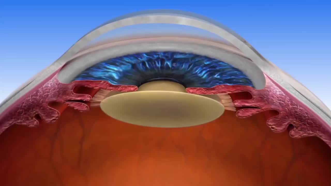
Glaucoma is a disease that damages the nerve at the back of your eye, called the optic nerve. The optic nerve sends the signals to the brain that enables you to see. Glaucoma is associated with increased sensitivity to pressure within the eye. The eye’s aqueous fluid is constantly produced and drained at a balanced rate and forms the intraocular pressure within the eye (IOP). When this drainage is reduced, or if there is too much fluid production, IOP increases and leads to damage of the optic nerve.


Image of the optic nerve showing loss of nerve cells in patients with glaucoma.
Glaucoma typically has no symptoms. Most patients do not experience any issues until they visit their eyecare provider. Untreated glaucoma can cause a gradual loss of vision, usually starting in the periphery which may go unnoticed for long periods of time.

Image showing the effect of loss of sight in patients with glaucoma.
What to expect :

Assessment
Your appointment will take between 30 to 90 mins, depending on the complexity of the consultations.
This includes:
- Questions on your medical and eye history (please bring a list of your medications and any relevant notes)
- Tests required to diagnose glaucoma which include measuring the pressure within the eye (tonometry), inspecting the drainage angle of the eye (gonioscopy), measuring the thickness of the cornea (pachymetry), assessing the function of your peripheral vision (visual fields) and detailed imaging of the nerve at back of the eye (OCT and photography).
- A dilated eye examination may be required. This involves using eye drops to make your pupil bigger and it takes some time for the drops to work. After dilation, your close-up vision may remain blurry for several hours, and you will be more sensitive to light. It is advisable not to drive for at least 2 hours after the drops have been instilled.

Meet your surgeon
We will perform a comprehensive examination of your eye and discuss your concerns.
Your treatment will be customised based on your tests and preferences.
Some options to help manage glaucoma include pressure lowering eye drops, gentle laser treatments and surgery in theatre.
Most patients are generally managed with daily prescription eye drops which decrease eye pressure by either reducing the production of fluid or increasing the outflow of fluid. The commonly prescribed eyedrops in New Zealand include Timolol, Betoptic, Combigan, Dortim, Lattim, Brimonidine, Latanoprost, Travoprost, Bimatoprost, Trusopt, Azopt, Pilocarpine and Iopidine.
It is important to take the medications as prescribed to you. Your family doctor can provide you with a repeat of these medications. Let us know if you have any side effects associated with glaucoma eye drops. Click here to watch a video on side effects.

Gentle Glaucoma Light Therapy (laser) in Clinic
There are various forms of laser treatments used in the treatment of glaucoma. For some patients, laser therapy can lower pressure and avoid the need for more invasive surgery later or reduce the need for medications or drops.
- Laser Peripheral Iridotomy and argon laser iridoplasty is used in the treatment of angle closure/narrow angle glaucoma.
- Selective Laser Trabeculoplasty is used in the treatment of open angle glaucoma.
These procedures are generally well tolerated and are done as a day procedure under topical anaesthesia (eyedrops). Recovery is excellent with most patients returning to work the following day.
Click these links for information on Laser Peripheral Iridotomy and Selective Laser Trabeculoplasty.

Glaucoma Surgery in Theatre
- Because there are several variables that lead to glaucoma surgery, procedures can last from 15 mins to 2 hours.
- Specific preoperative and post operative instructions will be provided, including a detailed discussion on potential risks to allow you to make informed consent.
- Following surgery, patients are kept comfortable for about an hour to ensure it was a success and to allow the sedative to wear off before they are released into the care of a family member or friend.
- Generally, patients do not experience significant pain after their procedure; however, they should expect their eye to feel irritated and their vision to be slightly blurred immediately after surgery. This is normal and will gradually improve over the days and weeks that follow.

Follow up
- Understand your medication routine and attend planned clinic follow up.
- As glaucoma typically has no symptoms, we can only assess the effect of the condition with tests. As such, regular lifetime follow up is crucial to prevent further vision loss.

5 most common reasons patients have LASIK eye surgery :
Improved Vision :
LASIK can significantly improve high definition vision, allowing people to see clearly without relying on glasses or contact lenses. This is a huge benefit for people who’ve been struggling with poor vision for years.
Convenience :
For those always on the go or have an active outdoor lifestyle, a life not limited by glasses or contacts is a huge lift.
Cost Savings :
Over time, the cost of glasses and/or contact lenses, all add up. LASIK is a very cost-effective solution to reduce long-term expenses related to vision correction.
Safety :
With advances in laser technology, the small risks associated with LASIK have become increasingly minimal. Risks are minimised by choosing an experienced specialist laser surgeon who uses the latest laser technology.
Improved Self-Confidence :
For many, wearing glasses or contact lenses can be a source of self-consciousness. LASIK helps to improve self-confidence and allow people to feel more comfortable in their own skin.
Saving money
The hidden costs: what could you save?

What could that money have done?
The $40,000 you'd potentially spend on glasses and contact lenses over the next 30 years could have given you 15 luxurious holidays.
Est - 15 Holidays

Average holiday cost: $2600
Calculation: 40,000 / $2600 = 15
Saving time
The hidden cost of wearing glasses or contact lenses.

Did you know?
The time you'd spend handling glasses and contact lenses over the next 30 years is enough to have taken you around our planet over 30 times!
Est- 526 hours

Cleaning: 3 minutes/day
Finding misplaced glasses: 2 minutes/day
Repair, shopping: 2 hours/year
Total: 526 hours (over 30 years)
Est- 1,012 hours

Inserting and Removing: 3 minutes/day
Cleaning: 2 minutes/day
Renewing & Check-ups: 2 hours/year
Total: 1,012 hours (over 30 years)
Saving planet
Reducing our carbon footprint

The environmental perspective
Over 30 years, using glasses and contact lenses contributes to 876lb of CO2 emissions. Opt for vision correction and take a stand for our planet!
Est- 876.6 CO2 emissions

Combined plastic waste:
0.525 kg (glasses)+65.7 kg (contact lenses)=66.225 kilograms over three decades.
Total: 66.225 kilograms of plastic×6=397.35 kilograms = 876Lb of CO2 emissions(over 30 years)
Invested in Your Glaucoma Care
You are in safe hands at Re:Vision. We are committed to using the world’s most advanced technology and techniques to halt glaucoma progression so you can live a life full of sight.
Some patients are not suitable for drops or laser therapy and require either cataract surgery, minimally invasive glaucoma surgery or complex glaucoma surgery.
[Read More About Minimally Invasive Glaucoma Surgery (MIGS)]
[Read More About Complex Glaucoma Surgery (Trabeculectomy and Drainage Tubes)]
[Read More About Cataract Surgery and Glaucoma]
[Link associated with Minimally Invasive Glaucoma Surgery (MIGS)]
Minimally invasive Glaucoma surgery (MIGS) marks a milestone in the advancement of glaucoma patient care.
MIGS are a form of glaucoma surgery which is associated with minimal incisions, and are generally associated with lower risks in restoring normal eye pressure.
Prior to MIGS, treatment options were limited to medications, laser and major glaucoma tube and filtration surgery. Now, with MIGS, our team at Re:Vision have more treatment options which benefits a patient with faster recovery , less surgical risks and less dependence on medications following surgery.
Our surgeon is a leader in MIGS surgery in NZ, with a wealth of experience with iStent -W, Kahook Dual Blade, Hydrus, XEN, Preserflo, micropulse and cyclodiode laser.
MIGS management include
1) Various treatment options and benefits which are patient centric and individualised
2) Evidence based therapy and
3) Promising outcomes for glaucoma patients.
MIGS can be performed as a standalone procedure or in conjunction with cataract surgery. There are different surgical approaches under the MIGS umbrella, but they generally are designed to allow more fluid to drain out of the eye and hence, reduce intraocular pressure.
- iStent
The iStent is the smallest medical device ever implanted into humans. The iStent decreases eye pressure by creating a pathway into the eye’s drainage system. The iStent was the first trabecular micro-bypass device approved by the FDA and is the most thoroughly-studied glaucoma device on the market.
Click here on a video on iStent
- Kahook Dual Blade
The Kahook Dual Blade goniotomy procedure involves removing a section of the trabecular meshwork (the part which is associated with the greatest resistance in fluid outlflow) and hence increasing drainage out of the eye.
[Click here on a video on KDB]
- Micropulse Laser Treatment
This procedure decreases the amount of fluid produced by the eye and increases fluid outflow. A probe is placed on the surface of the eye and the surgeon applies laser energy to part of the eye which controls the production of fluid in the eye.
- Preserflo Stent
The Preserflo drainage shunt is type of microshunt implant used to reduced intraocular pressure by creating a controlled pathway for the drainage of fluid out of the eye. It is made from a specialised biocompatible material called SIBS, which is known for its stability, flexibility and compatibility with the human body, hence minimising tissue inflammation.
[Click here on a video on Preserflo]
[Link associated with Trabeculectomy and Tube Shunts]
Trabeculectomy surgery has been done for more than 50 years, and involves making a separate channel for fluid to filter out of the eye.
A drainage tube is a device inserted into the eye, which acts as straw where fluid can access to drain out of the eye onto the outer coat of the eye. There are several drainage tubes available in NZ which include Molteno drainage tube, Paul drainage tube and Baerveldt drainage tube.
Generally, these surgeries are done once all others have been exhausted and have failed to stop the progression of glaucoma.
Click here on a video on Trabeculectomy
Click here on a video on Drainage Tube
Link associated with Glaucoma and Cataracts
Glaucoma is optic nerve damage due to increased pressure sensitivity within the eye while a cataract is a clouding of your natural lens within your eye.
In some patients, the hardening and change in the shape of the lens with the development of cataract, can potentiate the effects of glaucoma, such as angle closure glaucoma. In this situation, we may suggest having cataract surgery as a way to treat your glaucoma.
Some patients can combine glaucoma and cataract surgery (includes patients with open angle glaucoma). MIGS procedures in particular can be performed through the same incision as cataract surgery, providing an efficient way to address two problems at once.
Personalised Surgical Care
Some patients are not suitable for drops or laser therapy and require either cataract surgery, minimally invasive glaucoma surgery or complex glaucoma surgery.
Minimally invasive Glaucoma surgery (MIGS) marks a milestone in the advancement of glaucoma patient care. MIGS are a form of glaucoma surgery which is associated with minimal incisions, and are generally associated with lower risks in restoring normal eye pressure.
Prior to MIGS, treatment options were limited to medications, laser and major glaucoma tube and filtration surgery. Now, with MIGS, our team at Re:Vision have more treatment options which benefits a patient with faster recovery , less surgical risks and less dependence on medications following surgery.
Our surgeon is a leader in MIGS surgery in NZ, with a wealth of experience with iStent -W, Kahook Dual Blade, Hydrus, XEN, Preserflo, micropulse and cyclodiode laser.
MIGS management include:
1) Various treatment options and benefits which are patient centric and individualised
2) Evidence based therapy and
3) Promising outcomes for glaucoma patients.
MIGS can be performed as a standalone procedure or in conjunction with cataract surgery. There are different surgical approaches under the MIGS umbrella, but they generally are designed to allow more fluid to drain out of the eye and hence, reduce intraocular pressure.
a) iStent
The iStent is the smallest medical device ever implanted into humans. The iStent decreases eye pressure by creating a pathway into the eye’s drainage system. The iStent was the first trabecular micro-bypass device approved by the FDA and is the most thoroughly-studied glaucoma device on the market.
Click here to watch video on iStent
b) Kahook Dual Blade
The Kahook Dual Blade goniotomy procedure involves removing a section of the trabecular meshwork (the part which is associated with the greatest resistance in fluid outlflow) and hence increasing drainage out of the eye.
Click here for information on KDB
c) Micropulse Laser Treatment
This procedure decreases the amount of fluid produced by the eye and increases fluid outflow. A probe is placed on the surface of the eye and the surgeon applies laser energy to part of the eye which controls the production of fluid in the eye.
d) Preserflo Stent
The Preserflo drainage shunt is type of microshunt implant used to reduced intraocular pressure by creating a controlled pathway for the drainage of fluid out of the eye. It is made from a specialised biocompatible material called SIBS, which is known for its stability, flexibility and compatibility with the human body, hence minimising tissue inflammation.
Click here for information on Preserflo
Trabeculectomy surgery has been done for more than 50 years, and involves making a separate channel for fluid to filter out of the eye.
A drainage tube is a device inserted into the eye, which acts as straw where fluid can access to drain out of the eye onto the outer coat of the eye. There are several drainage tubes available in NZ which include Molteno drainage tube, Paul drainage tube and Baerveldt drainage tube.
Generally, these surgeries are done once all others have been exhausted and have failed to stop the progression of glaucoma.
Glaucoma is optic nerve damage due to increased pressure sensitivity within the eye while a cataract is a clouding of your natural lens within your eye.
In some patients, the hardening and change in the shape of the lens with the development of cataract, can potentiate the effects of glaucoma, such as angle closure glaucoma. In this situation, we may suggest having cataract surgery as a way to treat your glaucoma.
Some patients can combine glaucoma and cataract surgery (includes patients with open angle glaucoma). MIGS procedures in particular can be performed through the same incision as cataract surgery, providing an efficient way to address two problems at once.
World’s most advanced glaucoma machines
Triton Swept Source OCT
Triton Swept Source OCT (Optical Coherence Tomography) is a high-tech imaging system used in ophthalmology for detailed, non-invasive imaging of the eye. This technology utilises a Swept Source laser to capture cross-sectional images of the retina and other structures within the eye.
It provides high-resolution, three-dimensional images that allow eye care professionals to visualize and analyse various layers of the retina, optic nerve, and other structures with great detail. This level of detail is crucial in diagnosing and managing various eye conditions such as macular degeneration, diabetic retinopathy, and glaucoma.
The Triton Swept Source OCT is valued for its speed, accuracy, and ability to provide comprehensive imaging of the eye, aiding in the early detection and monitoring of eye diseases. It's considered a significant advancement in eye care technology, enhancing the ability of eye care professionals to assess and manage ocular health.

Humphrey Visual Field Analyser
The Humphrey Visual Field Analyzer (HFA) is a diagnostic tool used in ophthalmology to assess a person's visual field, which refers to the full extent of what can be seen peripherally while focusing on a central point. It's particularly valuable in detecting and monitoring conditions affecting the visual field, such as glaucoma, optic nerve damage, neurological disorders, or certain retinal diseases.
The HFA employs a computerized testing method to map out a person's visual field. During the test, the patient focuses on a fixed point while lights of varying intensity are presented in different areas of their peripheral vision. The patient signals when they detect these lights, and the machine records the responses to create a map of their visual field sensitivity.
This information is crucial in diagnosing and monitoring conditions that affect the visual field, especially in diseases like glaucoma where peripheral vision loss can occur gradually and without noticeable symptoms in the early stages. Regular HFA tests allow clinicians to track changes in the visual field over time, aiding in treatment decisions and assessing the progression of the condition.
The Humphrey Visual Field Analyzer is a widely used and respected tool in eye care due to its accuracy, reliability, and ability to provide quantitative data about a patient's visual field.

Ellex Tango Reflex Neo YAG/SLT machine
The Ellex Tango Reflex Neo SLT/YAG machine is a sophisticated medical device used in ophthalmology for treating glaucoma.
Selective laser trabeculoplasty involves using a laser to treat the drainage area of the eye (trabecular meshwork) responsible for regulating the eye's internal pressure by improving fluid outflow. The laser is applied in short, low-energy pulses to specific areas of the trabecular meshwork, aiming to increase drainage efficiency without causing significant damage to surrounding tissues.
Laser peripheral iridotomy is performed to treat angle closure glaucoma.

What is my glaucoma risk?

.webp)
Although anyone may develop glaucoma, people at greater risk include:
- A family history of glaucoma (first-degree relatives)
- High eye pressure (>21mmHg)
- Age over 50
- Thin cornea
- Diabetes
- Short or long sightedness
- Migraine
- High or low blood pressure
- Obstructive sleep apnoea
You can calculate your risks using the online calculator here
What is Wavefront-guided LASIK ?
.webp)
What is Wavefront-Guided LASIK?
Wavefront-Guided LASIK uses advanced scanning technology to map and correct 25 unique higher-order aberrations in your visual system—not just your glasses prescription. It provides a highly customised laser correction, leading to sharper, clearer vision and better outcomes in night vision and contrast sensitivity.
Why Choose Wavefront-Guided LASIK?
Wavefront-Guided LASIK is the safest, most precise laser vision correction method available today. It offers faster recovery, less discomfort, and clearer vision—especially in challenging conditions like night driving. It's also the most flexible option for future enhancements.
How is WG-LASIK Different from SMILE?
SMILE corrects only basic focus errors (like myopia or mild astigmatism) and cannot customise treatment to your unique eye structure. WG-LASIK uses highly detailed scans to treat subtle optical imperfections, resulting in better night vision, more accurate outcomes, and easier enhancement options later.
Is LASIK right for me?
Most people with stable vision, healthy corneas, and realistic expectations are suitable for LASIK. We’ll use 3D eye scans and wavefront diagnostics to determine if LASIK—or another treatment—will give you the best long-term result.
How long does the surgery take?
The procedure itself takes only 10–15 minutes per eye. You’ll be in the clinic for around 1.5–2 hours total, including prep and recovery time. Vision often improves within hours after surgery.
Does LASIK surgery hurt?
No—LASIK is virtually painless. We use anaesthetic drops to numb the eye. You may feel light pressure for a few seconds, but most patients describe the procedure as surprisingly comfortable.
When can I drive or return to work?
Most LASIK patients can drive and return to work within 1–2 days. Vision typically stabilises quickly, especially with wavefront-guided technology. We'll give you clear post-op guidance based on your specific case.
Can you guarantee me perfect vision with LASIK?
While no procedure can guarantee “perfect” vision, WG-LASIK offers the most accurate and predictable outcomes available. Most patients achieve 20/20 or better. If your vision changes in future, enhancement options are available.
Is there an upper age limit for LASIK?
There’s no strict upper age limit, but your suitability depends on your eye health, corneal thickness, and visual goals. Many people in their 40s, 50s, and beyond enjoy excellent outcomes with LASIK or other tailored procedures.
Do I need to leave contact lenses out before surgery?
Yes. Contact lenses can alter the shape of your cornea. We recommend leaving soft lenses out for at least 2–3 days, and rigid lenses out for longer, before your scans and surgery. We'll advise the exact timing during your assessment.
How long will the correction last?
Most patients enjoy stable vision for many years. WG-LASIK permanently reshapes the cornea, but natural age-related changes (like presbyopia) may still occur over time. Enhancement options are available if needed in the future.
The difference between LASIK and SMILE :
Here are reasons our surgeons at Re:Vision see LASIK as a superior procedure to SMILE:


Precision
LASIK is auto-centred; SMILE is manual and less accurate.

Vision Quality
LASIK offers high-definition vision; SMILE is less crisp.

Recovery Speed
LASIK recovers in days; SMILE takes weeks or longer.

Dryness Outcomes
Both procedures show similar dry eye symptoms by 3 months post-surgery.

Astigmatism Accuracy
LASIK corrects astigmatism more precisely than SMILE, thanks to auto-alignment.

Error Correction
LASIK treats 25 focus errors; SMILE treats only myopia and astigmatism.

Fine-Tuning
LASIK is easy to fine-tune; SMILE is harder and riskier.

Night Vision
LASIK provides better night vision thanks to a wider treatment zone and error correction

Surgeon Involvement
SMILE requires much more surgeon hand-held manipulation than LASIK.
Glaucoma Research at Re:Vision

Dr Perumal values the importance of research initiatives and technology in glaucoma. We may invite you to participate in the Save Sight Registry run by the Save Sight Institute, in collaboration with the University of Sydney and Sydney Eye Hospital to fight glaucoma blindness.
The Save Sight Registries is one of the most advanced ophthalmic registries in the world, and is a unique platform for tracking eye disease, interventions and patient outcomes. Its sophisticated design delivers real-world evidence on the risks and benefits of current and new treatments for ocular conditions. This information helps clinicians provide safe, cost-effective and evidence-based solutions for vision impairment and avoidable blindness.
The Save Sight Registries also promotes international scientific research aimed at developing strategies for reducing the incidence of blindness throughout the world.
Frequently Asked Questions:
What is glaucoma?
Glaucoma occurs when pressure inside the eye causes damage to the optic nerve, resulting in loss of peripheral vision, which can progress to central vision and lead to blindness.
Is glaucoma treatable or curable?
Glaucoma is not curable and unfortunately, the damage to your optic nerve is permanent. Glaucoma is, however, treatable. Treatment requires management of intraocular pressure to prevent further damage.
How do I know if I've got glaucoma?
Annual eye exams are key to knowing if you have glaucoma.
Detection involves accurate measurement of intraocular pressure and scans to establish the function and structure of the optic nerve.
What are the types of glaucoma?
Glaucoma refers to a group of diseases that cause damage to the optic nerve as a result of increased pressure sensitivity within the eye.
There are two main types of glaucoma:
- Open-angle glaucoma and Angle-closure glaucoma. Open-angle glaucoma is the most common type of glaucoma and involves fluid in the eye not draining properly through the trabecular meshwork.
- Angle-closure glaucoma involves a sudden buildup of pressure in the eye and poor drainage because the angle between the iris and the cornea is too narrow.
- Many patients do not experience any symptoms during the early stages of glaucoma, including no pain and no vision loss. This makes it difficult for many patients to know if they have the disease. But as glaucoma progresses, patients may experience a loss of peripheral or side vision, along with sudden eye pain, headache, blurred vision or the appearance of halos around lights.
- Other forms of glaucoma include secondary glaucoma (such as trauma, surgery, pigment dispersion, pseudoexfoliation glaucoma), congenital glaucoma (present from birth) and medication induced.
How do I prevent glaucoma?
Ophthalmologists are still researching the cause of glaucoma; however, early detection and appropriate management is crucial to prevent further damage.
Is glaucoma hereditary?
Glaucoma does have a genetic component so having a family member with this disease is a risk factor.
What is considered dangerously high eye pressure?
“Normal" eye pressure will vary from one person to the next, and in patients with pressure sensitivity, their eye pressure needs to be lower than a patient with normal visual function. However, an eye pressure above 21mmHg will need further investigations. Consult with your optometrist if you have any concerns.
Can glaucoma patients live a normal life?
Although patients can be nervous about a glaucoma diagnosis, it is important to keep a positive attitude. Glaucoma does not have to limit your lifestyle, and for the most part, you should be able to continue your regular habits and activities.
Can I have low eye pressure and still have glaucoma?
Glaucoma can still be present with low eye pressure.
What role do I play in the management of glaucoma?
You play an important role in the management of your glaucoma. It is important to follow the advice and adhere to the treatment plan developed for you.
Contact your family members to request glaucoma screening tests via their eyecare provider.
If you have any questions regarding glaucoma or your treatment, contact us directly.
Additional resources include
- Glaucoma NZ https://glaucoma.org.nz/
- Glaucoma Australia https://glaucoma.org.au/
- American Academy of Ophthalmology https://www.aao.org
Costs
At Re:Vision, all types of laser vision correction (LASIK or PRK) cost :$7,000
A detailed quote for your consultation will be provided prior to your consultation.
We will not charge you for any procedure unless discussed and your consent is obtained.


At Re:Vision, all types of laser vision correction (LASIK or PRK) cost : $7,600
We want to make paying for your treatment as easy as possible so we offer a range of finance options to help you get the outcome you want.
Your eyes are unique and we customise all of our treatments to give you the best vision possible.
You might be surprised to learn how much money you save when compared to a lifetime of buying glasses or contact lenses.

Premium laser eye surgery or lens implant surgery is an investment that you won’t regret.
See how much you can save with laser eye surgery
By the time I turn 45 I will have spent :
on contact lenses/glasses
24 month interest free finance options :
*Offer applies to QCARD & GEM finance. Lending criteria, fees & T&C'S apply.
*New Zealand residents only

Still have questions?
Get in touch with us and a friendly member of our team will assist.
References :


